")

Diagnostics
After taking the patients history, plain radiographs of the affected part of the spine are performed. However, x-ray pictures only depict the bony structures of the spine. Therefore in most cases cross section studies like Computed tomography (CT) and Magnetic resonance Imaging (MRI) are indispensable. Especially by means of MRI, soft tissue structures like nerves and intervertebral discs can be excellently illustrated without harmful x-ray exposure. For an exact differentiation of the pain´s origin, it is in some cases necessary to carry out diagnostic injections. Here a pinpoint local anesthesia is placed under x-ray control into the region of interest. During discography and myelography, anatomic structures of the spine are visualized by means of contrast media. These invasive diagnostics are still necessary despite the availability of CT and MRI, because both the discs and the spinal canal can be illustrated under strain. In addition, during these diagnostic steps a pain relieving medication can be added to the injection in order to reach a lasting therapeutic effect.
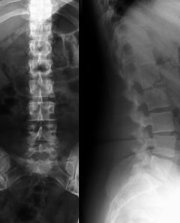
X-ray image of the lumbar spine
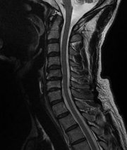
MRI image of the cervical spine
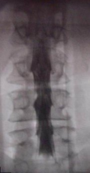
Myelogram of the lumbar spine
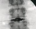
Discography of a lumbar vertebral disc